
Page 87 - วารสารปีที่15ฉบับที่2
P. 87
212
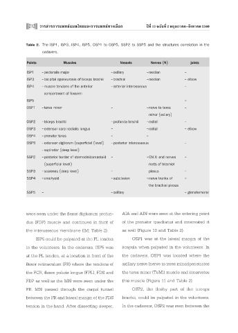
Table 2. The ISP1, ISP3, ISP4, ISP5, OSP1 to OSP5, SSP2 to SSP5 and the structures correlation in the
cadavers.
Points Muscles Vessels Nerves (N) joints
ISP1 -pectoralis major -axillary -median -
ISP3 -bicipital aponeurosis of biceps brachii -brachial -median - elbow
ISP4 -muscle tendons of the anterior -anterior interosseous -
compartment of forearm
ISP5 -
OSP1 -teres minor - -nerve to teres -
minor (axiiary)
OSP2 -triceps brachii -profunda brachii -radial -
OSP3 -extensor carpi radialis longus - -radial - elbow
OSP4 -pronator teres - -
OSP5 -extensor digitorum (superficial (level) -posterior interosseous -
-supinator (deep level)
SSP2 -posterior border of sternocleidomastoid - -CN XI and nerves -
(superficial level) roots of brachial
SSP3 -scalenes (deep level) - plexus -
SSP4 -omohyoid -subclavian -nerve trunks of -
the brachial plexus
SSP5 - -axillary - glenohemoral
were seen under the flexor digitorum profun- AIA and AIN were seen at the entering point
dus (FDP) muscle and continued in front of of the pronator quadratus and innervated it
the interosseous membrane (IM; Table 2). as well (Figure 10 and Table 2).
ISP5 could be palpated at the PL tendon OSP1 was at the lateral margin of the
in the volunteers. In the cadavers, ISP5 was scapula when palpated in the volunteers. In
at the PL tendon, at a location in front of the the cadavers, OSP1 was located where the
flexor retinaculum (FR) where the tendons of axillary nerve (nerve to teres minor)penetrates
the FCR, flexor policis longus (FPL), FDS and the teres minor (TeMi) muscle and innervates
FDP as well as the MN were seen under the this muscle (Figure 11 and Table 2).
FR. MN passed through the carpal tunnel OSP2, the fleshy part of the triceps
between the FR and lateral margin of the FDS brachii, could be palpated in the volunteers.
tendon in the hand. After dissecting deeper, In the cadavers, OSP2 was seen between the