
Page 88 - วารสารปีที่15ฉบับที่2
P. 88
213
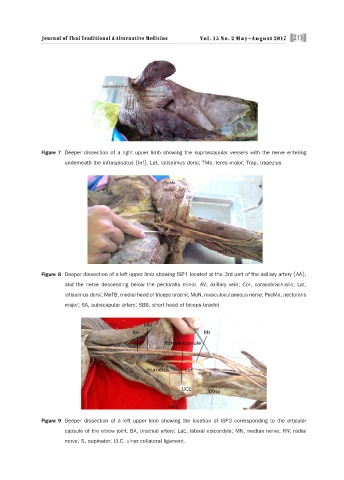
Figure 7 Deeper dissection of a right upper limb showing the suprascapular vessels with the nerve entering
underneath the infraspinatus (Inf). Lat, latissimus dorsi; TMa, teres major; Trap, trapezius
Figure 8 Deeper dissection of a left upper limb showing ISP1 located at the 3rd part of the axillary artery (AA),
and the nerve descending below the pectoralis minor. AV, axillary vein; Cor, coracobrachialis; Lat,
latissimus dorsi; MeTB, medial head of triceps brachii; MuN, musculocutaneous nerve; PecMa, pectoraris
major; SA, subscapular artery; SBB, short head of biceps brachii
Figure 9 Deeper dissection of a left upper limb showing the location of ISP3 corresponding to the articular
capsule of the elbow joint. BA, brachial artery; LaE, lateral epicondyle; MN, median nerve, RN; radial
nerve; S, supinator; ULC, ulnar collateral ligament.