
Page 89 - วารสารปีที่15ฉบับที่2
P. 89
214
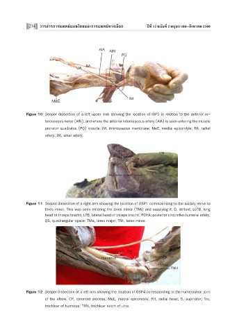
Figure 10 Deeper dissection of a left upper limb showing the location of ISP5 in relation to the anterior in-
terosseous nerve (AIN), and where the anterior interosseous artery (AIA) is seen entering the muscle
pronator quadratus (PQ) muscle. IM, interosseous membrane; MeE, medial epicondyle; RA, radial
artery; UA, ulnar artery.
Figure 11 Deeper dissection of a right arm showing the location of OSP1 corresponding to the axillary nerve to
teres minor. This was seen entering the teres minor (TMi) and supplying it. D, deltoid; LoTB, long
head of triceps brachii, LTB; lateral head of triceps brachii; PCHA, posterior circumflex humeral artery;
QS, quadrangular space; TMa, teres major; TMi, teres minor.
Figure 12 Deeper dissection of a left arm showing the location of OSP4 corresponding to the humeroulnar joint
of the elbow. CP, coronoid process; MeE, medial epicondyle; RH, radial head; S, supinator; Tro,
trochlear of humerus; TUN, trochlear notch of ulna.